Natural Product Efficacy Evaluation
Saposhnikovia divaricata root and its major components ameliorate inflammation and altered gut microbial diversity and compositions in DSS-induced colitis
Saposhnikovia divaricata root and its major components ameliorate inflammation and altered gut microbial diversity and compositions in DSS-induced colitis
- 저자: Erdenebileg S; Son YJ; Kim M; Oidovsambuu S; Cha KH; Kwon J; Jung DS; Nho CW
- 출처: - (2023)
- DOI/링크: https://doi.org/10.1016/j.imr.2023.100998
- 연구 계열: Topics/Natural Product Efficacy Evaluation
- 투고일: 2023-02-08
TL;DR
방풍(Saposhnikovia divaricata) 뿌리 메탄올 추출물(MESD)은 LPS로 자극한 RAW 264.7 세포에서 NO와 염증성 사이토카인을 유의하게 낮췄고, DSS 유도 대장염 마우스의 대장 염증성 사이토카인 발현도 줄였다. 장내 Clostridium sensu stricto 1 감소와 예측 기능 경로 변화도 관찰되었다.
1. 연구 배경 및 동기
- 다루는 문제: 방풍 뿌리는 항염·항산화·진통 성분을 가진 전통 약용식물이다. 그러나 염증성 장질환(IBD)에 대한 항염 효과를 과학적으로 검증한 연구는 없었다.
- 기존의 공백 / 왜 필요한가: 방풍 뿌리의 IBD 완화 효과와 장내미생물 변화를 확인한 연구가 없다.
2. 관련 연구
- 기존 연구 흐름: 방풍 뿌리와 성분이 RAW 264.7 세포에서 염증성 사이토카인, iNOS, COX-2를 억제하고, 일부 성분이 마우스 통증과 LPS 유도 IL-6·NO 생성을 억제한다는 보고가 있다.
- 이 논문의 위치: RAW 264.7 세포와 DSS 대장염 마우스 모델에서 항염 효과를 확인하고 16S rRNA 분석으로 장내미생물 변화를 함께 살핀 연구이다.
3. 핵심 기여
- MESD의 시험관 및 DSS 대장염 마우스에서의 항염 효과를 제시
- 장내미생물 조성 변화(Clostridium sensu stricto 1 감소)를 확인
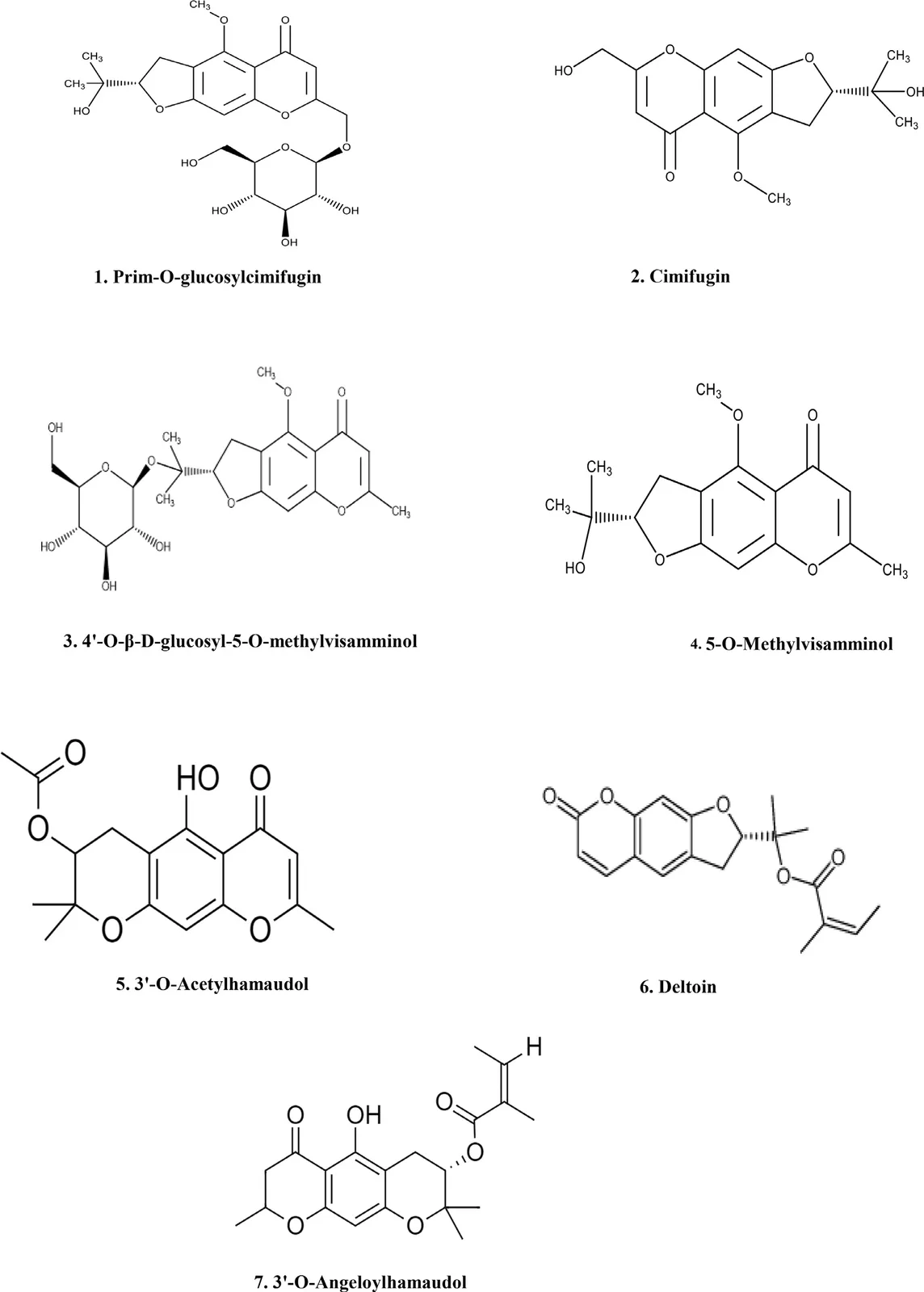
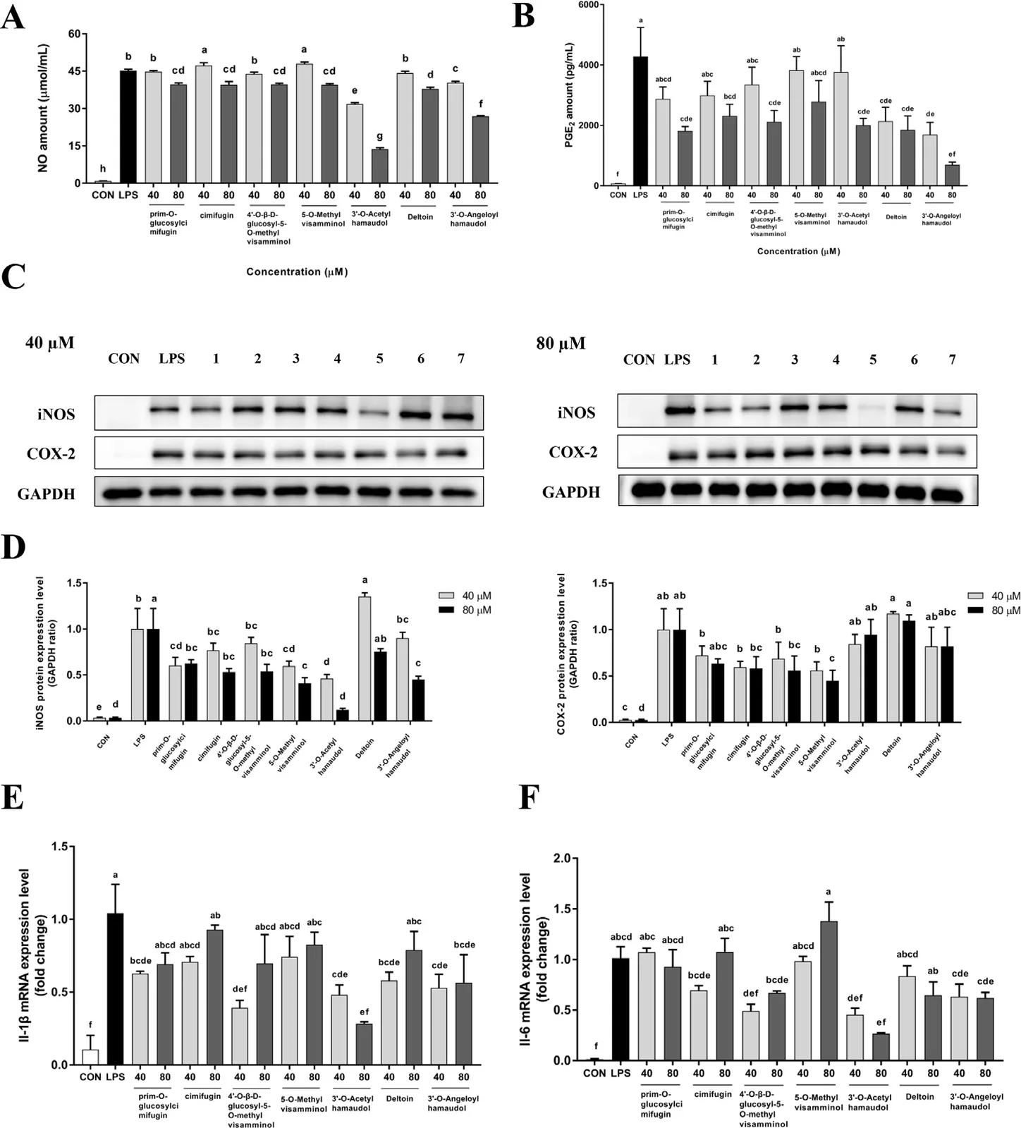
- 항염 활성 화합물 7종을 분리하고 3’-O-acetylhamaudol과 3’-O-angeloylhamaudol의 강한 시험관 항염 효과를 확인
4. 제안 방법론
- 전체 접근 논리: 추출물의 항염 효과를 세포와 DSS 대장염 모델에서 검증하고, 16S rRNA 서열분석으로 장내미생물 변화를 평가한 뒤, 추출물에서 활성 성분을 분리했다.
- 단계:
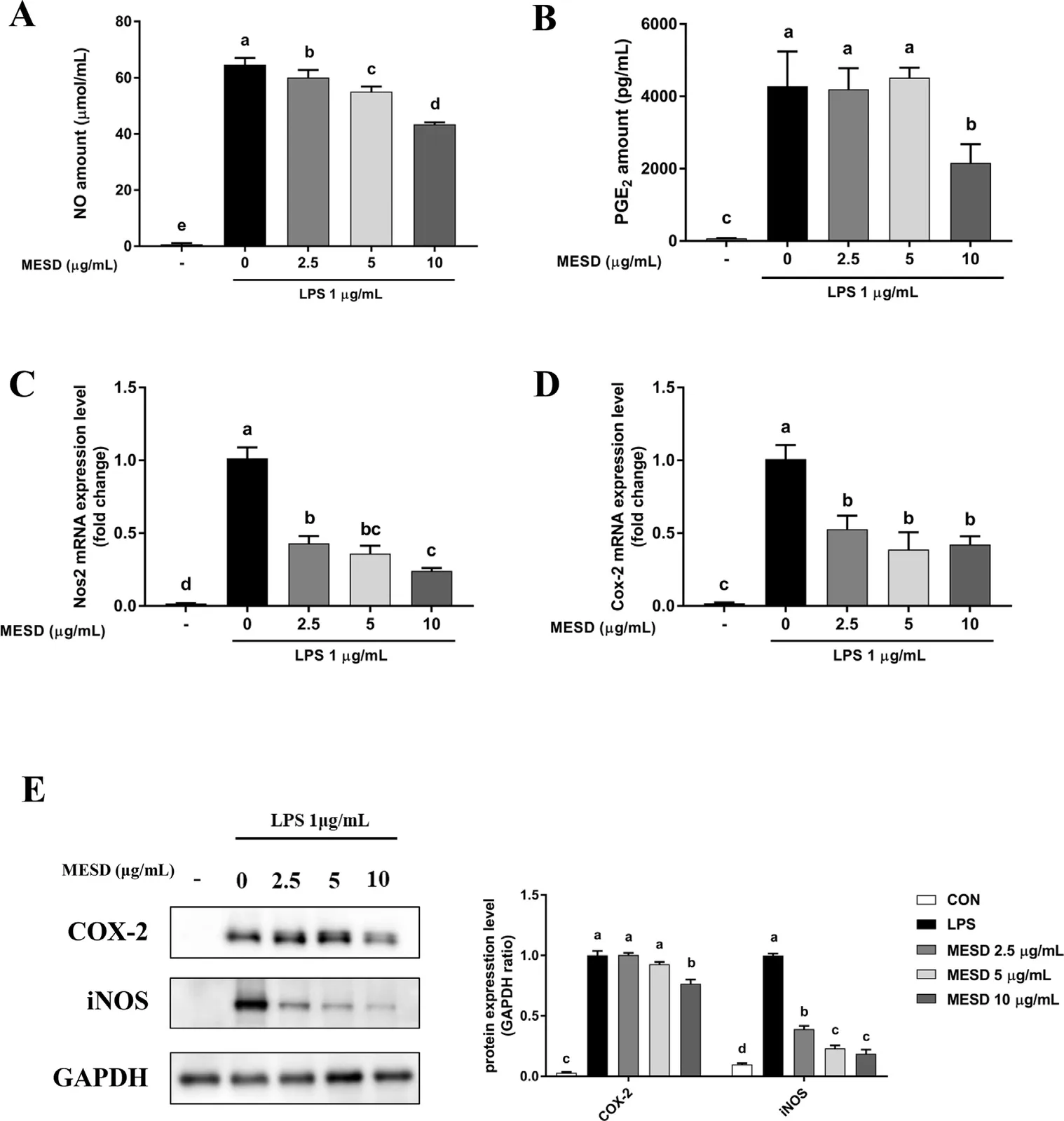
- MESD 처리 후 LPS 유도 RAW 264.7 세포의 NO와 사이토카인 측정
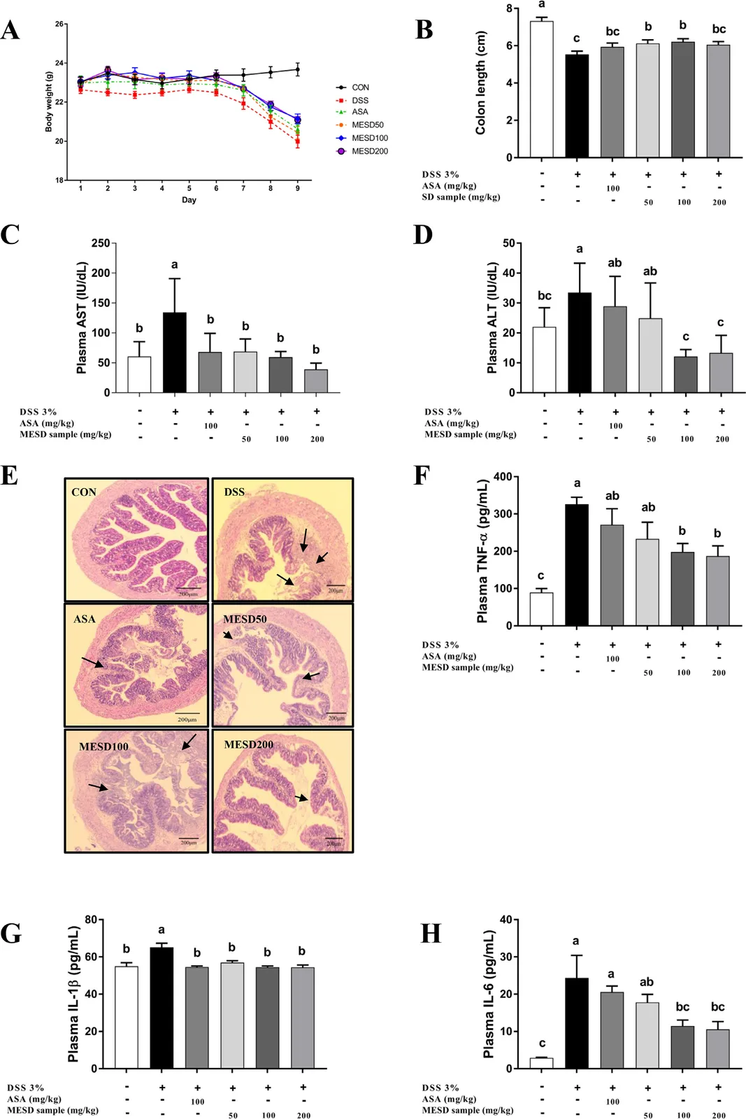
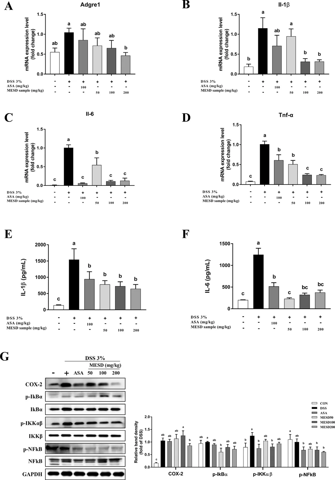
- DSS 유도 대장염 마우스에 MESD 경구 투여 후 대장 염증성 사이토카인 발현 분석
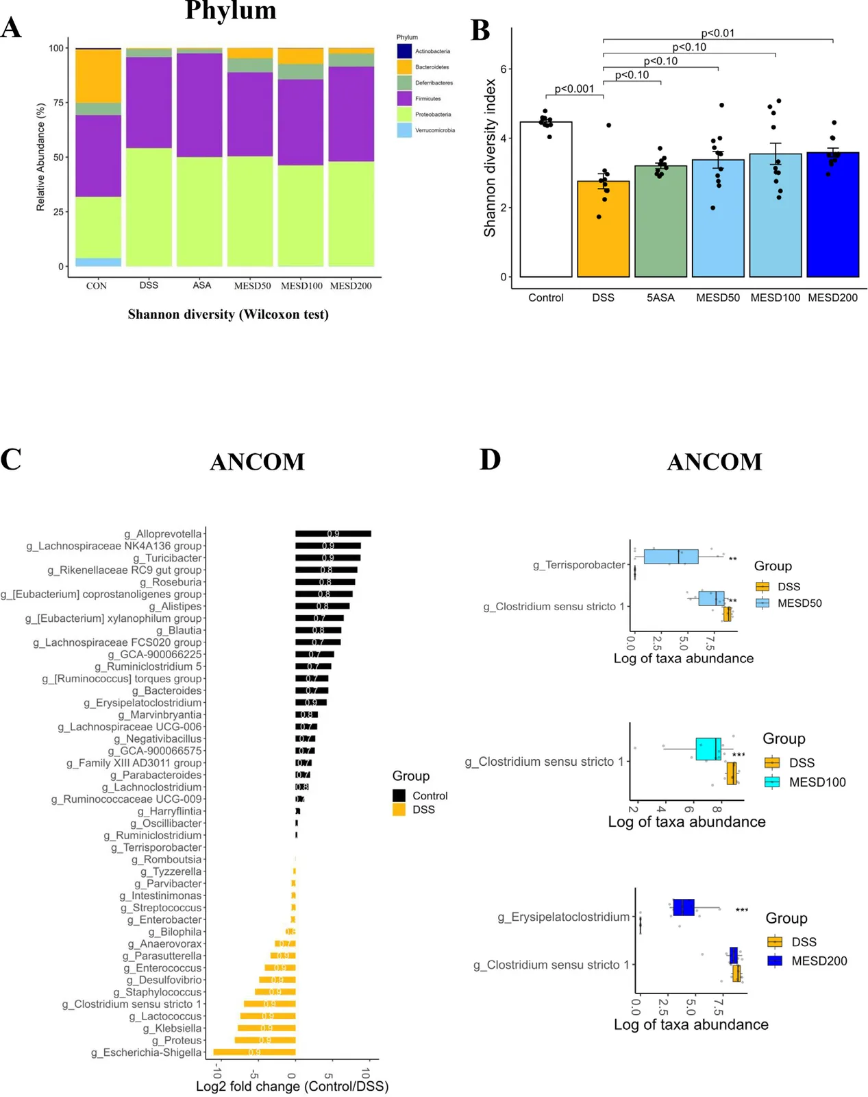
- 16S rRNA 유전자 서열분석으로 장내미생물 변화 확인
- MESD에서 항염 성분 7종 분리, 시험관 활성 평가
5. 실험 설정
| 항목 | 내용 |
|---|---|
| 대상/재료 | RAW 264.7 마우스 대식세포, DSS 유도 대장염 마우스 |
| 처리 조건 | 방풍 뿌리 메탄올 추출물(MESD) 처리, 분리 화합물 처리 |
| 대조군 | - |
| 측정 방법 | NO, 염증성 사이토카인, 16S rRNA 서열분석, 예측 기능 경로 |
6. 실험 결과 분석
| 지표 | 변화(Δ) | 조건 · 유의성 |
|---|---|---|
| NO 및 염증성 사이토카인 | 유의하게 감소 | LPS 유도 RAW 264.7 세포, MESD |
| 대장 염증성 사이토카인 발현 | 감소 | DSS 대장염 마우스, MESD 경구 투여 |
| Clostridium sensu stricto 1 존재량 | 억제 | MESD, 16S rRNA 분석 |
| 예측 기능 경로 | L-glutamate degradation VIII(프로피온산 생성) 등이 증강 | MESD |
| 분리 화합물 | 항염 성분 7종, 그중 3’-O-acetylhamaudol과 3’-O-angeloylhamaudol이 시험관에서 강한 항염 효과 | 시험관 |
제안 기전(인과 사슬):
MESD → 염증성 사이토카인 감소 및 장내미생물 조성 변화(Clostridium sensu stricto 1 감소) → 대장염 완화
7. 비판적 평가
강점
- 세포·동물·미생물 분석과 성분 분리를 함께 수행
- 활성 화합물을 특정
한계 · 의문
- 미생물 기능 경로는 예측 결과
- 분리 성분의 효과는 시험관 수준에서 확인
8. 향후 연구 방향
- MESD와 활성 성분의 IBD 치료 잠재력에 대한 추가 검증
9. 결론
MESD는 염증성 사이토카인을 낮추고 장내미생물 조성을 바꾸어 IBD 치료용 천연물이 될 가능성이 있다.
Keywords
Saposhnikovia divaricata, DSS-induced colitis, inflammatory bowel disease, gut microbiota, anti-inflammatory, hamaudol, RAW 264.7
원문 그림
원문 그림 · 라이선스: cc-by-nc-nd